« Back to General Dermatology FAQs

Diagnostic Techniques

» What is the most sensitive office laboratory test for diagnosing dermatophyte infections of the skin?
» How is a KOH examination performed?
» What laboratory tests are useful for diagnosing tinea capitis?
» What is a Wood’s light or lamp? How is it useful in skin diseases?
» Name common culture media used for isolating dermatophytes.
» Describe a simple test for tinea versicolor other than a KOH preparation.
» What is a Tzanck preparation or smear?
» What is the best method of diagnosing scabies?
» How do you diagnose mite bites acquired from an animal?
» How do you diagnose lice infestation?
» What is the diagnostic test of choice for a patient presenting with a suspected syphilitic chancre on his penis?
» How is secondary syphilis diagnosed?
» How long do serologic tests for syphilis remain positive?
» In patients with symptomatic gonococcal urethritis, how efficacious is a Gram stain of the exudate in comparison to a culture utilizing selective media for gonococcus?
» What is the best way to diagnose allergic contact dermatitis?
» How are patch tests applied?
» In what diseases is a skin biopsy helpful?
» When are shave biopsies indicated?
» What are the indications for punch biopsies?
» Describe the indications for an excisional or incisional biopsy.
» Define and describe direct immunofluorescence of the skin.
» Name some skin diseases in which DIF is helpful in making a diagnosis.
» How does indirect immunofluorescence of the skin differ from direct immunofluorescence of the skin?
» Is ELISA ever used for the diagnosis of immunobullous disease?
» How are bacterial skin cultures performed, and when are they useful?


Name some skin diseases in which DIF is helpful in making a diagnosis.


Direct immunofluorescent study demonstrating granular deposits of IgA within a blood vessel of a patient with Henoch- Schönlein purpura. (Courtesy of James E. Fitzpatrick, MD.)
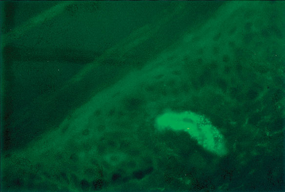
Fig. 3.8 Direct immunofluorescent study demonstrating granular deposits of IgA within a blood vessel of a patient with Henoch- Schönlein purpura. (Courtesy of James E. Fitzpatrick, MD.)
Many of the immunobullous diseases are associated with specific DIF findings: bullous pemphigoid, herpes gestationis, cicatricial pemphigoid, epidermolysis bullosa acquisita, dermatitis herpetiformis, linear IgA bullous dermatosis, and the various types of pemphigus. In addition, DIF may be helpful in evaluating cutaneous and systemic lupus erythematosus, other collagen vascular diseases, vasculitis such as Henoch-Schönlein purpura (Fig. 3-8), and certain types of porphyria.