« Back to General Dermatology FAQs

Diagnostic Techniques

» What is the most sensitive office laboratory test for diagnosing dermatophyte infections of the skin?
» How is a KOH examination performed?
» What laboratory tests are useful for diagnosing tinea capitis?
» What is a Wood’s light or lamp? How is it useful in skin diseases?
» Name common culture media used for isolating dermatophytes.
» Describe a simple test for tinea versicolor other than a KOH preparation.
» What is a Tzanck preparation or smear?
» What is the best method of diagnosing scabies?
» How do you diagnose mite bites acquired from an animal?
» How do you diagnose lice infestation?
» What is the diagnostic test of choice for a patient presenting with a suspected syphilitic chancre on his penis?
» How is secondary syphilis diagnosed?
» How long do serologic tests for syphilis remain positive?
» In patients with symptomatic gonococcal urethritis, how efficacious is a Gram stain of the exudate in comparison to a culture utilizing selective media for gonococcus?
» What is the best way to diagnose allergic contact dermatitis?
» How are patch tests applied?
» In what diseases is a skin biopsy helpful?
» When are shave biopsies indicated?
» What are the indications for punch biopsies?
» Describe the indications for an excisional or incisional biopsy.
» Define and describe direct immunofluorescence of the skin.
» Name some skin diseases in which DIF is helpful in making a diagnosis.
» How does indirect immunofluorescence of the skin differ from direct immunofluorescence of the skin?
» Is ELISA ever used for the diagnosis of immunobullous disease?
» How are bacterial skin cultures performed, and when are they useful?


What is the best method of diagnosing scabies?


A positive scraping for scabies showing an immature mite, eggs, and numerous fecal pellets. (Courtesy of James E. Fitzpatrick, MD.)
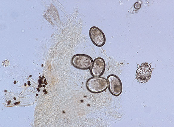
Fig. 3.6 A positive scraping for scabies showing an immature mite, eggs, and numerous fecal pellets. (Courtesy of James E. Fitzpatrick, MD.)
The best method is to scrape a burrow and demonstrate the parasite inside it. A classic burrow appears as an irregular, linear, slightly elevated lesion, best found on the flexor wrists, fingerwebs, and genitalia. Eighty-five percent of adult male patients with scabies will have mites on the hands or wrists. Occasionally, the mite can be seen with the naked eye as a small dot at one end of the burrow. Following the application of mineral oil, on either the blade or skin,  the burrow is scraped vigorously with a no. 15 scalpel blade, but not so vigorously as to draw blood. The mineral oil is collected from the skin and blade and transferred to a glass slide, which is then examined under the microscope. The diagnosis is established by identifying either the fecal pellets (scybala), eggs, or the mite itself (Fig. 3-6).
Hay RJ: Scabies and pyodermas—diagnosis and treatment, Dermatol Ther 22:466–464, 2009.