« Back to General Dermatology FAQs

Diagnostic Techniques

» What is the most sensitive office laboratory test for diagnosing dermatophyte infections of the skin?
» How is a KOH examination performed?
» What laboratory tests are useful for diagnosing tinea capitis?
» What is a Wood’s light or lamp? How is it useful in skin diseases?
» Name common culture media used for isolating dermatophytes.
» Describe a simple test for tinea versicolor other than a KOH preparation.
» What is a Tzanck preparation or smear?
» What is the best method of diagnosing scabies?
» How do you diagnose mite bites acquired from an animal?
» How do you diagnose lice infestation?
» What is the diagnostic test of choice for a patient presenting with a suspected syphilitic chancre on his penis?
» How is secondary syphilis diagnosed?
» How long do serologic tests for syphilis remain positive?
» In patients with symptomatic gonococcal urethritis, how efficacious is a Gram stain of the exudate in comparison to a culture utilizing selective media for gonococcus?
» What is the best way to diagnose allergic contact dermatitis?
» How are patch tests applied?
» In what diseases is a skin biopsy helpful?
» When are shave biopsies indicated?
» What are the indications for punch biopsies?
» Describe the indications for an excisional or incisional biopsy.
» Define and describe direct immunofluorescence of the skin.
» Name some skin diseases in which DIF is helpful in making a diagnosis.
» How does indirect immunofluorescence of the skin differ from direct immunofluorescence of the skin?
» Is ELISA ever used for the diagnosis of immunobullous disease?
» How are bacterial skin cultures performed, and when are they useful?


Describe a simple test for tinea versicolor other than a KOH preparation


A positive cellophane tape preparation of tinea versicolor that has been stained with methylene blue. The characteristic clusters of spores and short hyphae are demonstrated. (Courtesy of James E. Fitzpatrick, MD.)
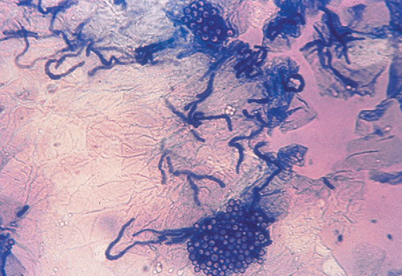
Fig. 3.4 A positive cellophane tape preparation of tinea versicolor that has been stained with methylene blue. The characteristic clusters of spores and short hyphae are demonstrated. (Courtesy of James E. Fitzpatrick, MD.)
Clear cellophane tape preparations are an excellent diagnostic testing material because the organism is found in the upper stratum corneum. First, the skin is scraped to ensure there is adequate scale. The tape is applied over the scale and then mounted on a glass slide and examined under the microscope. Clusters of short hyphae and yeasts are seen producing a “spaghetti and meatballs” pattern. Methylene blue may also be added to the slide, selectively staining the organism and thus enhancing visualization (Fig. 3-4). It is important to note that Malassezia globosa and M. furfur, the most common etiologic agents of tinea versicolor, cannot be cultured on any of the routine fungal media kept in most laboratories.

Martin AG, Kobayashi GS: Yeast infections: candidiasis, pityriasis (tinea) versicolor. In Fitzpatrick TB, Eisen AZ, Wolff K, et al, editors: Dermatology in general medicine, ed 4, New York, 1993, McGraw-Hill, pp 2462–2467.