« Back to General Dermatology FAQs

Diagnostic Techniques

» What is the most sensitive office laboratory test for diagnosing dermatophyte infections of the skin?
» How is a KOH examination performed?
» What laboratory tests are useful for diagnosing tinea capitis?
» What is a Wood’s light or lamp? How is it useful in skin diseases?
» Name common culture media used for isolating dermatophytes.
» Describe a simple test for tinea versicolor other than a KOH preparation.
» What is a Tzanck preparation or smear?
» What is the best method of diagnosing scabies?
» How do you diagnose mite bites acquired from an animal?
» How do you diagnose lice infestation?
» What is the diagnostic test of choice for a patient presenting with a suspected syphilitic chancre on his penis?
» How is secondary syphilis diagnosed?
» How long do serologic tests for syphilis remain positive?
» In patients with symptomatic gonococcal urethritis, how efficacious is a Gram stain of the exudate in comparison to a culture utilizing selective media for gonococcus?
» What is the best way to diagnose allergic contact dermatitis?
» How are patch tests applied?
» In what diseases is a skin biopsy helpful?
» When are shave biopsies indicated?
» What are the indications for punch biopsies?
» Describe the indications for an excisional or incisional biopsy.
» Define and describe direct immunofluorescence of the skin.
» Name some skin diseases in which DIF is helpful in making a diagnosis.
» How does indirect immunofluorescence of the skin differ from direct immunofluorescence of the skin?
» Is ELISA ever used for the diagnosis of immunobullous disease?
» How are bacterial skin cultures performed, and when are they useful?


How is a KOH examination performed?


Refractile and cylindrical hyphae traversing KOH preparation of skin scraping.
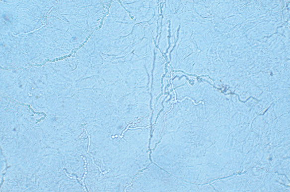
Fig. 3.1 Refractile and cylindrical hyphae traversing KOH preparation of skin scraping.
The highest rate of recovery of organisms occurs in specimens taken from the tops of vesicles, the leading edges of annular lesions, or deep scrapings from the nails suspected to be infected with fungi. The site should be swabbed with an alcohol pad or water and scraped with a no. 15 blade. In some instances, such as the scalp or nail, a curette may be more effective. The moist corneocytes are then easily transferred from the blade to a glass slide. One or two drops of KOH (10% to 20%) are added, and a coverslip is applied to the specimen. The KOH preparation is gently warmed, but not boiled, and then examined under the microscope. It is important to focus back and forth through the material so that the refractile hyphae can be visualized. Fungal hyphae can be recognized by their regular cylindrical shapes with branching and the presence of septae that may demonstrate a subtle greenish hue (Fig. 3-1). A pencil eraser or pen cap gently applied to the surface of the coverslip may enhance keratinocyte breakdown, especially for clumped specimens. If no organisms are observed initially, waiting 10 to 15 minutes may aid visualization.