« Back to General Dermatology FAQs

Diagnostic Techniques

» What is the most sensitive office laboratory test for diagnosing dermatophyte infections of the skin?
» How is a KOH examination performed?
» What laboratory tests are useful for diagnosing tinea capitis?
» What is a Wood’s light or lamp? How is it useful in skin diseases?
» Name common culture media used for isolating dermatophytes.
» Describe a simple test for tinea versicolor other than a KOH preparation.
» What is a Tzanck preparation or smear?
» What is the best method of diagnosing scabies?
» How do you diagnose mite bites acquired from an animal?
» How do you diagnose lice infestation?
» What is the diagnostic test of choice for a patient presenting with a suspected syphilitic chancre on his penis?
» How is secondary syphilis diagnosed?
» How long do serologic tests for syphilis remain positive?
» In patients with symptomatic gonococcal urethritis, how efficacious is a Gram stain of the exudate in comparison to a culture utilizing selective media for gonococcus?
» What is the best way to diagnose allergic contact dermatitis?
» How are patch tests applied?
» In what diseases is a skin biopsy helpful?
» When are shave biopsies indicated?
» What are the indications for punch biopsies?
» Describe the indications for an excisional or incisional biopsy.
» Define and describe direct immunofluorescence of the skin.
» Name some skin diseases in which DIF is helpful in making a diagnosis.
» How does indirect immunofluorescence of the skin differ from direct immunofluorescence of the skin?
» Is ELISA ever used for the diagnosis of immunobullous disease?
» How are bacterial skin cultures performed, and when are they useful?


What is a Wood’s light or lamp? How is it useful in skin diseases?


Wood’s light examination of the groin area demonstrating classic coral red fluorescence associated with erythrasma. (Courtesy of John L. Aeling, MD.)
Fig. 3.2 Wood’s light examination of the groin area demonstrating classic coral red fluorescence associated with erythrasma. (Courtesy of John L. Aeling, MD.)
Wood’s light examination of the urine in a patient with porphyria cutanea tarda demonstrating classic coral red fluorescence with normal urine specimen exhibited for comparison. (Courtesy of James E. Fitzpatrick, MD.)
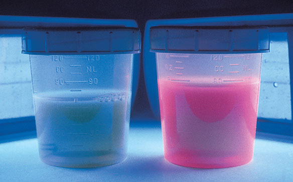
Fig. 3.3 Wood’s light examination of the urine in a patient with porphyria cutanea tarda demonstrating classic coral red fluorescence with normal urine specimen exhibited for comparison. (Courtesy of James E. Fitzpatrick, MD.)
A Wood’s light produces invisible long-wave ultraviolet radiation, or “black light,” at a wavelength of 360 nm. When this light strikes the surface of the skin or urine, fluorescence is produced in some disorders. This fluorescence is best observed in a completely dark room. The Wood’s lamp is useful in diagnosing cases of several skin conditions: tinea capitis (see preceding text), tinea versicolor (dull yellow fluorescence), erythrasma (coral red fluorescence; Fig. 3-2), and Pseudomonas infections of the skin (green fluorescence). It is also useful as a screening test in porphyria cutanea tarda as the urine fluoresces a coral red color (Fig. 3-3). The Wood’s light may also be used in certain disorders of pigmentation. In patients with hyperpigmentation, it is used to localize the site of the pigment because it accentuates superficial epidermal pigment, whereas deeper dermal pigment is unchanged. It is also used in patients with vitiligo because it demonstrates complete depigmentation. Finally, it can be used to delineate the borders of melanocytic lesions, such as lentigo maligna, prior to surgery.