« Back to General Dermatology FAQs

Diagnostic Techniques

» What is the most sensitive office laboratory test for diagnosing dermatophyte infections of the skin?
» How is a KOH examination performed?
» What laboratory tests are useful for diagnosing tinea capitis?
» What is a Wood’s light or lamp? How is it useful in skin diseases?
» Name common culture media used for isolating dermatophytes.
» Describe a simple test for tinea versicolor other than a KOH preparation.
» What is a Tzanck preparation or smear?
» What is the best method of diagnosing scabies?
» How do you diagnose mite bites acquired from an animal?
» How do you diagnose lice infestation?
» What is the diagnostic test of choice for a patient presenting with a suspected syphilitic chancre on his penis?
» How is secondary syphilis diagnosed?
» How long do serologic tests for syphilis remain positive?
» In patients with symptomatic gonococcal urethritis, how efficacious is a Gram stain of the exudate in comparison to a culture utilizing selective media for gonococcus?
» What is the best way to diagnose allergic contact dermatitis?
» How are patch tests applied?
» In what diseases is a skin biopsy helpful?
» When are shave biopsies indicated?
» What are the indications for punch biopsies?
» Describe the indications for an excisional or incisional biopsy.
» Define and describe direct immunofluorescence of the skin.
» Name some skin diseases in which DIF is helpful in making a diagnosis.
» How does indirect immunofluorescence of the skin differ from direct immunofluorescence of the skin?
» Is ELISA ever used for the diagnosis of immunobullous disease?
» How are bacterial skin cultures performed, and when are they useful?


What is a Tzanck preparation or smear?


A positive Tzanck preparation demonstrating large multinucleated keratinocytes. The nuclei of normal keratinocytes are the size of neutrophils, which are the other cells present in this preparation. (Courtesy of James E. Fitzpatrick, MD.)
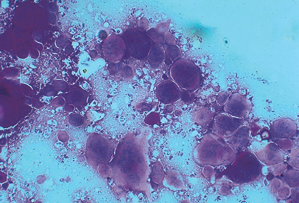
Fig. 3.5 A positive Tzanck preparation demonstrating large multinucleated keratinocytes. The nuclei of normal keratinocytes are the size of neutrophils, which are the other cells present in this preparation. (Courtesy of James E. Fitzpatrick, MD.)
A Tzanck smear is a standard technique for the rapid diagnosis of herpes simplex virus (HSV) or varicellazoster virus (VZV) infections. It cannot distinguish between these two agents, nor can it distinguish between HSV subtypes (HSV type 1 or 2). It is performed by scraping the base of a fresh blister with a scalpel blade and then spreading the adhering cells and material onto a glass slide. The slide is then stained with a Giemsa, Wright, or Sedi stain. The typical multinucleated giant cells or atypical keratinocytes with large nuclei are then easily visualized (Fig. 3-5).

Nahass GT, Goldstein BA, Zu WY, et al: Comparison of Tzanck smear, viral culture, and DNA diagnostic methods in detection of herpes simplex virus and varicella-zoster infection, JAMA 268:2541–2544, 1992.