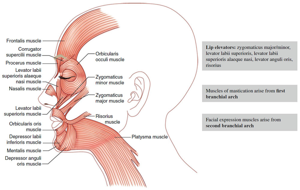
| Table 6-1 Muscles of Head and Neck |
| | Muscle | | Function | | Innervation |
| | Occipitalis | | Pulls scalp posteriorly | | Posterior auricular br. of facial nerve (CN VII)
{Do not confuse with postauricular branch of trigeminal nerve (CNV)} |
| | Frontalis muscle | | Elevates eyebrows and wrinkles forehead (horizontal forehead lines) | | Temporal br. of CN VII |
| | Orbicularis oculi | | Blinking and tight closure of eyelids (“crow’s feet”), lesser role as brow depressor (depressor supercilii) | | Temporal br. of CN VII (upper portion), zygomatic branch of CN VII (lower portion) |
| | Corrugator supercilii | | Pulls eyebrows medially and downward (vertical glabellar lines) | | Temporal br. of CN VII |
| | Procerus | | Pulls medial portion of eyebrows and glabellar skin downward (horizontal glabellar lines over root of nose) | | Zygomatic and buccal br. of CN VII per Bolognia (rare sources say temporal br.) |
| | Nasalis | | Alar flaring and compression
(“bunny lines” over upper bridge of nose) | | Zygomatic and buccal br. of CN VII |
| | Levator labii superioris | | Elevates upper lip | | Buccal br. of CN VII |
| | Levator labii superioris alaeque nasi | | Lifts upper lip, dilates nostrils | | Buccal br. of CN VII |
| | Levator anguli oris | | Lifts corners of the mouth | | Buccal br. of CN VII |
| | Risorius | | Produces smile by drawing back corners of mouth | | Marginal mandibular br. of CN VII per Bolognia (other sources say buccal br.) |
| | Zygomaticus major | | Main contributor to smile: elevates and draws corner of mouth laterally | | Buccal br. of CN VII |
| | Zygomaticus minor | | Elevates upper lip | | Buccal br. of CN VII |
| | Modiolus | | Accounts for cheek dimples in some patients | | |
| | Orbicularis oris | | Closes and purses lips
(vertical perioral lip lines) | | Buccal or marginal mandibular br. of CN VII |
| | Buccinator | | Presses cheek against teeth, allows blowing of cheeks | | Buccal br. of CN VII |
| | Depressor anguli oris | | Pulls corner of mouth downward (marionette lines → vertical lines at oral commissure) | | Marginal mandibular (MM) br. per Bolognia (most other sources say both MM and buccal br.) |
| | Depressor labii inferioris | | Depresses lower lip | | Marginal mandibular br. CN VII |
| | Mentalis | | Protrudes lower lip | | Marginal mandibular br. CN VII |
| | Platysma | | Pulls corner of mouth inferiorly, tenses neck (horizontal neck lines) | | Marginal mandibular br (upper portion) and cervical br. CN VII |
| | | | | | |