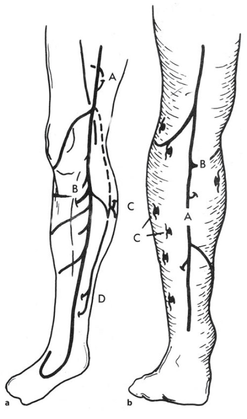
Perforating Veins | Fig. 8.3. a Typical course of the
long saphenous vein (LSV),
including its common tributaries
and perforating veins.
A Hunterian perforating vein,
B posterior tibial perforating
vein, C calf perforator in the
location of the intrasaphenous
vein,D medial ankle, or Cockett
perforators. b Typical course of
the SSV with termination above
the popliteal fossae and
associated perforator veins.
A SSV, B intersaphenous vein
with calf perforator,
C paraperoneal perforating veins.
(Reprinted with permission from
Goldman MP (1991)
Sclerotherapy: Treatment of
varicose and telangiectatic leg
veins. Mosby, St. Louis.) | Perforating/communicating veins connect the veins of the superficial venous system to the deep venous system by directing the one-way flow of blood into the deep venous system (Fig. 8.2). The only exception to this inward flow of blood is in the foot, where perforating veins with valves allow blood flow from the deep to the superficial veins [2]. Additionally, the majority of pedal-perforating veins usually do not contain valves. Therefore, a muscle pump in the foot must also provide a mechanism for venous return, which is activated by weight bearing. Perforating veins are present from the ankle to the groin. The number of perforating veins per leg is variable, with as few as 64 to as many as 155 per leg [2].Most perforating veins contain one to three valves.The valvular system is unidirectional,with blood flowing from superficial veins to deep veins. This prevents the high venous pressure from muscle contractions of the deep venous system from being transmitted to the superficial veins (Fig. 8.1). The typical perforating vein is a 1- to 2-mm thin-walled vessel. When perforating veins become incompetent, the high pressure from the deep veins of the calf-muscle pump is transmitted to the superficial veins by way of the perforating veins.When incompetent, perforating veins become thick-walled and may reach a diameter of 5 mm of more. In the thigh, the Hunterian (or Dodd’s) perforating veins are relatively constant and are associated with the medial intramuscular septum of the thigh (Fig. 8.2). These perforating veins connect the long saphenous vein to the femoral vein in the middle medial thigh and the lower third of the thigh. These perforating veins do not pierce muscle and consequently lose the benefit of protection from becoming incompetent by lack of support from the surrounding thigh muscles within the deep fascial compartment [3]. Hence, incompetence of the Hunterian/Dodd’s perforating veins is a common cause for medial thigh varicosities in patients with a competent saphenofemoral junction.Many smaller perforating veins may also be present in the middle third of the lateral aspect of the thigh and the midline posterior thigh, connecting the long saphenous vein and its tributaries to the profunda femoris vein (deep femoral vein).
The posterior tibial perforating vein occurs in almost all limbs approximately 5–10 cm distal to the knee on the medial calf (Fig. 8.3). It connects the long saphenous vein to the posterior tibial vein. Cockett’s perforating veins are comprised of a group of perforating veins located along the medial ankle coursing superiorly to the medical calf (Figs. 8.2 and 8.3). They do not drain directly into the long saphenous vein but connect the posterior arch vein of the calf to the posterior tibial vein (Figs. 8.2 and 8.3). Multiple perforating veins are also found with regularity along the medial calf. Some of these perforating veins drain into the posterior tibial vein, the gastrocnemius vein, and the soleal vein. Boyd’s perforating vein is another clinically important perforating vein, located approximately 10 cm below the medial joint of the knee. Perforating veins, therefore, play a fundamental roll in the development of varicose veins.
|