The Deep Venous System
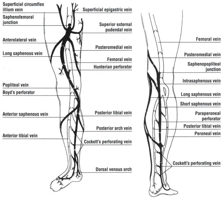
The principle veins of the deep venous system of the lower extremity consist of the anterior tibial, the posterior tibial and the peroneal veins, named for their corresponding paired arteries. These veins originate in the foot as plantar digital veins. At the level of the knee, these three veins join into a single popliteal vein (Fig. 8.2). The popliteal vein becomes the femoral vein (sometimes called the superficial femoral vein) once within the thigh (Fig. 8.2). The deep femoral vein (also referred to as the profunda femoris vein) joins the superficial femoral vein of the deep venous system proximally to form the common femoral vein. | ||
© 2025 Skin Disease & Care | All Rights Reserved.