Venous Anatomy of the Lower Extremity
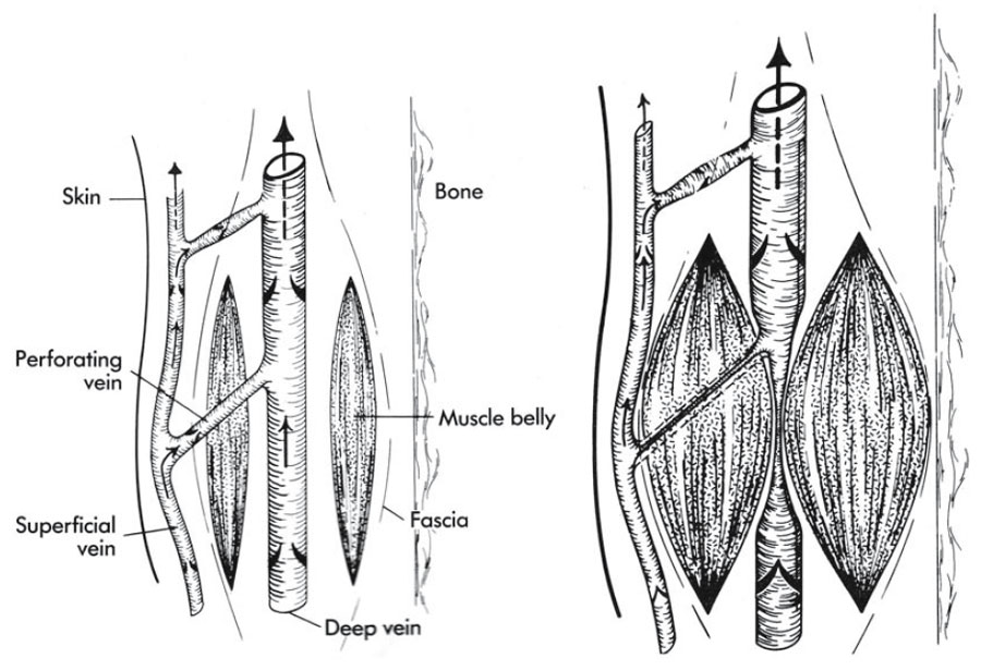
| Fig. 8.1a,b. Schematic diagram
of the calf-muscle pump. a
Relaxed state: all valves are open
allowing blood to flow in a
proximal direction. Blood flows
proximal in both the superficial
vein and through the perforating
vein into the deep veins. bWith
muscle contraction, the
perforating veins are squeezed
closed. Valves distal to the
compression are closed to
prevent distal blood flow.
(Reprinted with permission from
Goldman MP (1991)
Sclerotherapy: Treatment of
varicose and telangiectatic
leg veins. Mosby, St. Louis.) | An understanding of the venous system of the lower leg is important for proper treatment of varicose and telangiectatic veins. The venous systems of the leg are divided into two systems: deep and superficial. These two systems run parallel to the long axis of the leg. Ninety percent of the deoxygenated blood returning from the lower extremities is carried by the veins of the deep venous system [3]. The main function of the vessels of the superficial venous system is drainage of the venules of the skin into the deep venous system. The deep and superficial venous systems directly communicate through a series of perforating veins and also at venous junctions,where the blood of the superficial venous system drains into the deep venous system [4]. The veins of the deep venous system lie within the muscular system of the leg deep in its fascial compartment. The superficial veins course through the skin and subcutaneous tissue peripheral to the deep fascia. The perforating veins run an oblique course through the deep fascia, between muscle bundles, connecting the two systems. The mechanism of transporting venous blood from the legs is accomplished by contraction of the calf muscles (the calf-muscle pump or the peripheral heart) [2]. Therefore, the deep venous system is an important component in maintaining function of the cardiovascular system. During muscle relaxation, deep venous blood reflux in the leg is prevented by means of a passive oneway valve system (Fig. 8.1). Blood flow from the superficial to the deep venous system occurs via the perforating veins and the venous junctions during muscle relaxation when the hydrostatic pressure in the deep venous system falls below the pressure in the superficial veins (Fig. 8.1). The hydrostatic pressure in the saphenous veins of the lower extremities can be as high as 90–120 mmHg or more at the ankle when standing erect and motionless [2]. In contrast, the hydrostatic pressure at the distal aspect of the upper extremity at rest and upright is only 35 mmHg [5]. Pressure generated in the deep venous system can reach 200–300 mmHg during calf-muscle contraction, such as with walking (Fig. 8.1). Clearly, any source of prolonged increased hydrostatic pressure, such as prolonged standing or sitting in one place, will adversely affect the effectiveness of the calfmuscle pump.
|