« Back to Inflammatory Disorders

Deposition Disorders

»How is “deposition disorder” defined?
»What is amyloid?
»How is amyloid identified?
»Name the various types of amyloidosis.
»What are the cutaneous manifestations of primary or myeloma-associated systemic amyloidosis? How often do they occur?
»Name the other organ systems that may be involved in primary or myeloma-associated amyloidosis.
»Compare lichen amyloidosis and macular amyloidosis.
»How does nodular amyloidosis present? With what is it associated?
»In what setting is secondary systemic amyloidosis seen?
»What are the systemic manifestations of secondary systemic amyloidosis?
»What is lipoid proteinosis?
»What is colloid milium?
»Which histologic feature or “deposit” is common to all porphyrias?
»Which porphyria classically demonstrates the largest deposits? What are its cutaneous features?
»Name some of the cutaneous mucinoses.
»Describe the clinical lesions seen in pretibial myxedema and its disease associations.
»Describe the clinical lesions seen in lichen myxedematosus.
»What serum abnormality has been associated with scleromyxedema?
»Describe the clinical lesions in scleredema and its disease associations.
»What is a digital mucous (myxoid) cyst?
»What substance is elevated in gout?
»Where is the uric acid deposited in gout? What are the resulting clinical manifestations?
»How is gout treated?
»How many types of calcinosis cutis are there?
»What underlying medical conditions have been associated with metastatic calcinosis cutis?
»What is calciphylaxis and who develops it?
»What is osteoma cutis?


How is amyloid identified?

Lichen amyloidosis. Thioflavin-T demonstrates strong staining of fluorescent amyloid in the papillary dermis. (Courtesy of James E. Fitzpatrick, MD.)
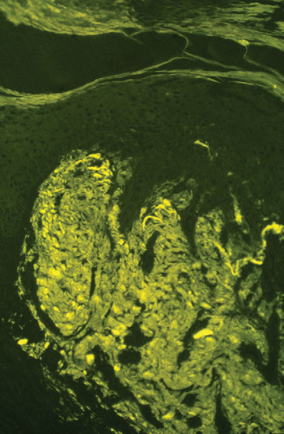
Fig. 16.1 Lichen amyloidosis. Thioflavin-T demonstrates strong staining of fluorescent amyloid in the papillary dermis. (Courtesy of James E. Fitzpatrick, MD.)
With light microscopy, amyloid appears as amorphous, hyaline-like, eosinophilic deposits. Amyloid demonstrates green birefringence with the alkaline Congo red stain, reddish metachromasia with crystal violet, and yellow-green fluorescence with thioflavin-T stain (Fig. 16-1). These stains are not absolutely specific for amyloid, as false-positive results may occur with the other hyaline-like deposition disorders.