Toggle navigation
Skin Diseases
An Intro on Skin Diseases
The Anatomy of the Skin
Some General Observations
Classification of the Elementary Lesions
Etiology
Local Dermal Inflammations
The Eruptions of Acute Specific Diseases
Papular Inflammations
Eczema
Bullous Diseases and Anomalous forms
Suppurative Inflammation
Squamous Inflammation
Diathetic Diseases
Hypertrophic and Atrophic Affections
Ichthyosis
Scleriasis
Formations or Neoplasmata
Cutaneous Haemorrhages
Pruritus
Chromatogenous diseases (alteration in the pigmentation)
Parasitic diseases
Favus
Disorders of the glands
Lichen Propicus or Prickly Heat
Diseases of the hair and hair follicle
Various lesions not classified
Dermatology
An Intro on Dermatology
Basic Science and Immunology
Melanocytes, Langherhans & Merkel Cells
Hair and Nails
Pediatric Dermatology
Childhood Infectious Diseases
Bullous Diseases
Tumors of Fat, Muscle and Bone
Genodermatoses
Syndromes with premature aging
Dermal Disorders
Diseases of the hair and nails
General Dermatology
Papulosquamous, Lichenoid & Eczematous
Granulomatous Diseases
Erythemas and Purpuras
Vesiculobullous Diseases
Disorders of Fat
Pigmentary Disorders & Vitamin Defects
Disorders of Hair
Infectious Diseases
Bacterial Infections
Fungal Infections
Protozoa and Worms
Infestations
Benign and Malignant Tumors
Premalignant and Malignant Tumors
Dermatologic Surgery
Excisions, Flaps, and Grafts
Surgical Complications
Cryosurgery and Electrosurgery
Sutures, Antiseptics, and Dressings
Nail Surgery
Pharmacology and Drug Reactions
Immunosuppressant Drugs
Other Drugs in Skin Disease and Care
Drug Reactions and Interactions
Pathology
Dermoscopy and Electron Microscopy
Life After Boards
High Yield Facts and Buzz Words
Skin Care
An Intro on Skin Care
Basic about Skin
Biology of the Skin
Assessment and Planning Care
Protecting the skin and preventing breakdown
Emollients
Psychological and social aspects of skin care
Helping patients make the most of their treatment
Illness Managment : Psoriasis
Illness Managment : Eczema
Illness Managment : Acne
Skin cancer and its prevention
Infective skin conditions and infestations
Less common skin conditions
Cosmetic Dermatology
An Intro on Cosmetic Dermatology
Anti-Aging Medicine As It Relates to Dermatology
Hormonal Regulation of Aging
Oral Antioxidant Nutrients
Anti-Aging Skin Care Ingredient Technologies
Photoaging & Pigmentary Changes in Skin
Chemexfoliation & Superficial Resurfacing
Medium-Depth Chemical Peeling
Deep Chemical Peeling
Botulinum Toxin
Soft Tissue Augmentation
Laser Skin Resurfacing
Sclerotherapy
Sclerotherapy Techniques for the Treatment of Varicose Veins
Dermatology FAQs
An Intro to Dermatology FAQs
Top 100 Undisclosed Facts
General FAQs
Inherited Disorders
Inflammatory Disorders
Infections and Infestations
Cutaneous Manifestations of Internal Diseases
Benign Tumors of the Skin
Malignant Tumors of the Skin
Treatment of Skin Disorders
Special Patient Populations
Emergencies and Miscellaneous Problems
Content
»
Hair and Nails
»
Hair
»
Nails
»
Wound Healing and Cytokines
»
Immunology
»
Noncellular Component
»
Toll-Like Receptors (TLR)
»
Complement System
»
Cells of The Immune System
»
B Cells
»
T Cells
»
NK Cells
»
Mononuclear Phagocytes
»
Neutrophils
»
Eosinophils
»
Mast Cells
»
Langerhans Cells
»
Antibodies
»
Major Histocompatibility Complex
»
References
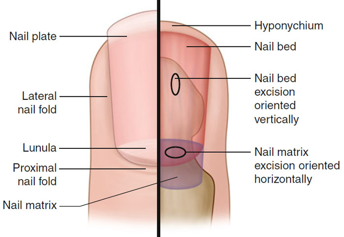
Nails
Figure 1.6
Nail anatomy
Nail plate
Consists of fully cornified cells (onychocytes); created by the nail matrix epithelium
Proximal nail matrix synthesizes the dorsal aspect of nail plate; distal nail matrix creates the ventral surface of the nail plate
Pink color of nail plate due to longitudinally situated subungual capillaries
Nail plate has firm attachment to underlying nail bed
Cuticle or eponychium: prevents separation of nail plate and proximal nail fold
Nail matrix:
Wedge-shaped area of specialized epithelium, divided into proximal and distal portion
Lunula demarcates distal portion of nail matrix
Melanocytes found in high concentration in nail matrix (mainly seen in the distal matrix)
Growth rate of fingernails 2–3 mm/month; toenails 1 mm/month
Complete replacement of nail requires 6 months for fingernail and 18 months for toenail
Skin Care
Skin Diseases
Dermatology
Cosmetic Dermatology
Dermatology FAQs
Home
Feedback
Disclaimer
© 2024 Skin Disease & Care | All Rights Reserved.