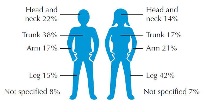
Type of melanoma Melanomas are categorised according to clinical and pathological parameters into four main types: superficial spreading melanoma, nodular melanoma, acral lentiginous melanoma and lentigo maligna melanoma. Superficial spreading melanoma is the most common type, accounting for approximately 70% (Cancer Research UK, 2009c). This type often grows slowly over a period of months or years and approximately 50% of patients will give a history of a pre-existing apparently benign lesion. The classic clinical presenting features include an irregular lateral margin, irregular multicoloured central pigmentation and a history of growth (Mackie, 2000). They are commonly found on the trunk in men and the leg in women, and patients are often in their fourth or fifth decade of life. The second most common type is nodular melanoma. These often present as raised lesions which bleed easily and are ulcerated. Although often dark in colour, they may be colourless or amelanotic. Lentigo maligna melanoma tends to occur on the face of elderly patients with extensive chronic sun damage. Lentigo maligna melanoma should not be confused with lentigo maligna, a type of melanoma in situ. Acral lentiginous melanoma, whilst rare, is the most common type of melanoma seen in Asians and people with dark skin. It is often found on the palms, soles, under fingernails and toenails and can affect mucous membranes. Risk factors Melanoma occurs most commonly in fairskinned persons, especially those with a history of significant sun exposure. Whilst sun exposure is thought to be a risk factor for melanoma, the relationship is not straightforward. The exact cause of melanoma is not known. Patients often have freckles, red hair and tan poorly. Having multiple naevi is a powerful predictor of risk of melanoma. Cutaneous melanoma can occur anywhere on the skin. Most frequently it is found on the leg in women (particularly between the knee and ankle) and on the trunk (especially the back) in men. In elderly people, melanomas develop most commonly on the face (Austoker, 1994). Figure 11.7 depicts the distribution of melanoma on parts of the body by sex.
| ||
© 2024 Skin Disease & Care | All Rights Reserved.