« Back to Inflammatory Disorders

Vesiculobullous Disorders

»What is the difference between a vesicle and a bulla?
»How are the bullous diseases defined?
»What things cause vesicles and bullae?
»How do you approach a patient who presents with an acute onset of a vesiculobullous eruption?
»Which skin findings are helpful in evaluating a patient with blisters?
»Do particular vesiculobullous diseases occur in characteristic distributions?
»Which tests are most useful in evaluating vesiculobullous diseases?
»How should a skin biopsy of a vesiculobullous eruption be performed?
»When are special tests necessary to diagnose blistering diseases of the skin?
»How are specimens obtained for direct immunofluorescence?
»For which vesiculobullous diseases are indirect immunofluorescence helpful?
»List the most common blistering diseases due to external agents.
»Name examples of drugs that can cause vesiculobullous eruptions.
»What is epidermolysis bullosa?
»Describe the other genetic blistering diseases.
»List the vesiculobullous diseases caused by metabolic disorders.
»Describe the clinical findings in bullous diabeticorum.
»What is the cause of pellagra?
»What is the difference between porphyria cutanea tarda and pseudoporphyria?
»What are the necrolytic erythemas?
»What is the difference between bullous pemphigoid and cicatricial pemphigoid?
»How do pemphigus vulgaris and pemphigus foliaceus differ?
»Linear IgA bullous dermatosis occurs in two different clinical situations. What are they?
»Describe the clinical findings in dermatitis herpetiformis.
»Does herpes gestationis have anything to do with herpes viruses?
»What is bullous systemic lupus erythematosus?
»What is epidermolysis bullosa acquisita?


Describe the clinical findings in dermatitis herpetiformis.

Dermatitis herpetiformis. Typical grouped vesicles and excoriations. The lesions are so intensely pruritic that in some patients only excoriations are seen.  (Courtesy of the Fitzsimons Army Medical Center teaching files.)
Fig. 10.6 Dermatitis herpetiformis. Typical grouped vesicles and excoriations. The lesions are so intensely pruritic that in some patients only excoriations are seen. (Courtesy of the Fitzsimons Army Medical Center teaching files.)
Direct immunofluorescence of skin demonstrating linear granular IgA along the basement membrane zone and in the papillary dermis in a patient with dermatitis herpetiformis.  (Courtesy of the Fitzsimons Army Medical Center teaching files.)
Fig. 10.2 Direct immunofluorescence of skin demonstrating linear granular IgA along the basement membrane zone and in the papillary dermis in a patient with dermatitis herpetiformis. (Courtesy of the Fitzsimons Army Medical Center teaching files.)
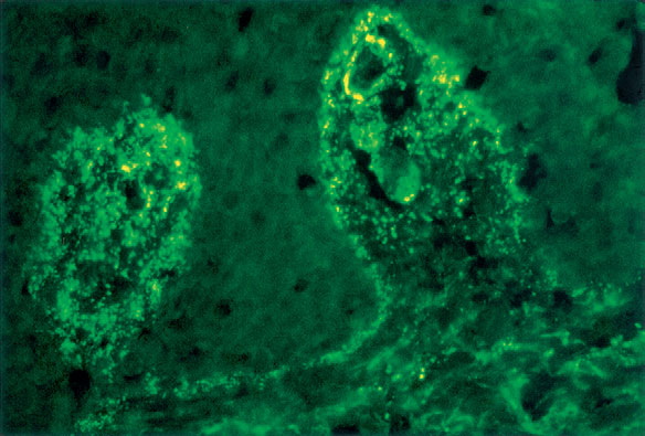
Dermatitis herpetiformis is an autoimmune disease due to IgA autoantibodies directed against tissue transglutaminase. Dermatitis herpetiformis, an extremely pruritic condition, most commonly begins in early adult life and is characterized by symmetrically distributed papules and vesicles that develop on the elbows, knees, buttocks, extensor forearms, scalp, and, sometimes, face and palms (Fig. 10-6). In some patients, the lesions are generalized and severe. Patients may have an associated gluten-sensitive enteropathy, though it is seldom symptomatic. The diagnosis is established by routine histologic exam of an early blister and direct immunofluorescence of nonlesional skin (IgA is seen in the dermal papillae) (see Fig. 10-2). Scratching may destroy all intact blisters for skin biopsy, and thus direct immunofluorescence may be a particularly helpful diagnostic test. Serologic testing for antiendomysial/eTG antibodies and antigliadin antibodies is also a useful screening test.

Alonso-Llamazares J, Gibson LE, Rogers RS 3rd: Clinical, pathologic, and immunopathologic features of dermatitis herpetiformis: review of the Mayo Clinic experience. Int J Dermatol 46:910-919, 2007.