« Back to Inflammatory Disorders

Vesiculobullous Disorders

»What is the difference between a vesicle and a bulla?
»How are the bullous diseases defined?
»What things cause vesicles and bullae?
»How do you approach a patient who presents with an acute onset of a vesiculobullous eruption?
»Which skin findings are helpful in evaluating a patient with blisters?
»Do particular vesiculobullous diseases occur in characteristic distributions?
»Which tests are most useful in evaluating vesiculobullous diseases?
»How should a skin biopsy of a vesiculobullous eruption be performed?
»When are special tests necessary to diagnose blistering diseases of the skin?
»How are specimens obtained for direct immunofluorescence?
»For which vesiculobullous diseases are indirect immunofluorescence helpful?
»List the most common blistering diseases due to external agents.
»Name examples of drugs that can cause vesiculobullous eruptions.
»What is epidermolysis bullosa?
»Describe the other genetic blistering diseases.
»List the vesiculobullous diseases caused by metabolic disorders.
»Describe the clinical findings in bullous diabeticorum.
»What is the cause of pellagra?
»What is the difference between porphyria cutanea tarda and pseudoporphyria?
»What are the necrolytic erythemas?
»What is the difference between bullous pemphigoid and cicatricial pemphigoid?
»How do pemphigus vulgaris and pemphigus foliaceus differ?
»Linear IgA bullous dermatosis occurs in two different clinical situations. What are they?
»Describe the clinical findings in dermatitis herpetiformis.
»Does herpes gestationis have anything to do with herpes viruses?
»What is bullous systemic lupus erythematosus?
»What is epidermolysis bullosa acquisita?


When are special tests necessary to diagnose blistering diseases of the skin?


Direct immunofluorescence of skin demonstrating linear granular IgA along the basement membrane zone and in the papillary dermis in a patient with dermatitis herpetiformis.  (Courtesy of the Fitzsimons Army Medical Center teaching files.)
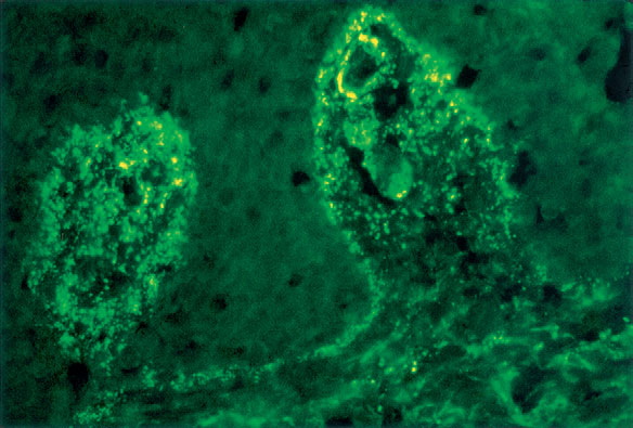
Fig. 10.2 Direct immunofluorescence of skin demonstrating linear granular IgA along the basement membrane zone and in the papillary dermis in a patient with dermatitis herpetiformis. (Courtesy of the Fitzsimons Army Medical Center teaching files.)
In addition to routine histology, a skin biopsy for direct immunofluorescence is often helpful in diagnosing the immunobullous diseases (Table 10-4). Direct immunofluorescent technique uses fluorescent, tagged antibodies that are directed against IgG, IgA, IgM, C3, and fibrin; these antibodies fluoresce when illuminated with a fluorescent microscope (Fig. 10-2). For precise diagnosis of the inherited forms of epidermolysis bullosa, electron microscopy studies may be necessary. Other tests are indicated in specific circumstances, such as urine porphyrin tests when porphyria cutanea tarda is being considered, and zinc levels when acrodermatitis enteropathica is possible.

Zillikens D: Diagnosis of autoimmune bullous skin diseases, Clin Lab 54:491–503, 2008.


Table 10-4. Direct Immunofluorescence Findings of Vesiculobullous Diseases
  DISEASE TARGET ANTIGEN DIRECT IMMUNOFLUORESCENCE
FINDINGS
 
Bullous pemphigoid
BP180, BP230
Linear C3, IgG at DEJ
 
Bullous SLE
COL7A1
Linear/granular IgG, other Igs at DEJ
 
Cicatricial pemphigoid
BP180, LAM5, and others
Linear C3, IgG, IgA at DEJ
 
Dermatitis herpetiformis
eTG
Granular IgA, C3 in upper dermis
(see Fig. 10-1)
 
Epidermolysis bullosa acquisita
COL7A1
Linear IgG, IgA, other Igs at DEJ
 
Herpes gestationis
BP180
Linear C3, IgG at DEJ
 
Linear IgA bullous dermatosis
BP180, COL7A1, LAD
Linear IgA, C3 at DEJ
 
Pemphigus foliaceus
DSG1
IgG, C3 in intercellular spaces
 
Pemphigus vulgaris
DSG3 (mucous membrane only)
DSG3 and DSG1 (mucous membrane and skin)
IgG, C3 in intercellular spaces
 
IgA pemphigus
DSC1, DSG1, DSG3
IgA in intercellular spaces
 
Paraneoplastic pemphigus
DSG1, DSG3, DP1, DP2, BP180, BP230, EP, PP, γ-catenin, plectin, 170 kD, DSC2, DSC3
IgG, C3 in intercellular spaces, DEJ
 
Porphyria cutanea tarda
None (not antibody mediated)
Homogenous IgG at DEJ and around vessels
 
Anti–p200 pemphigoid
200-kD antigen
IgG, C3 at DEJ
 
Anti–p105 pemphigoid
105-kD antigen
IgG, C3 at DEJ
           
DEJ, Dermal–epidermal junction; Ig, immunoglobulin; C3, third complement component.