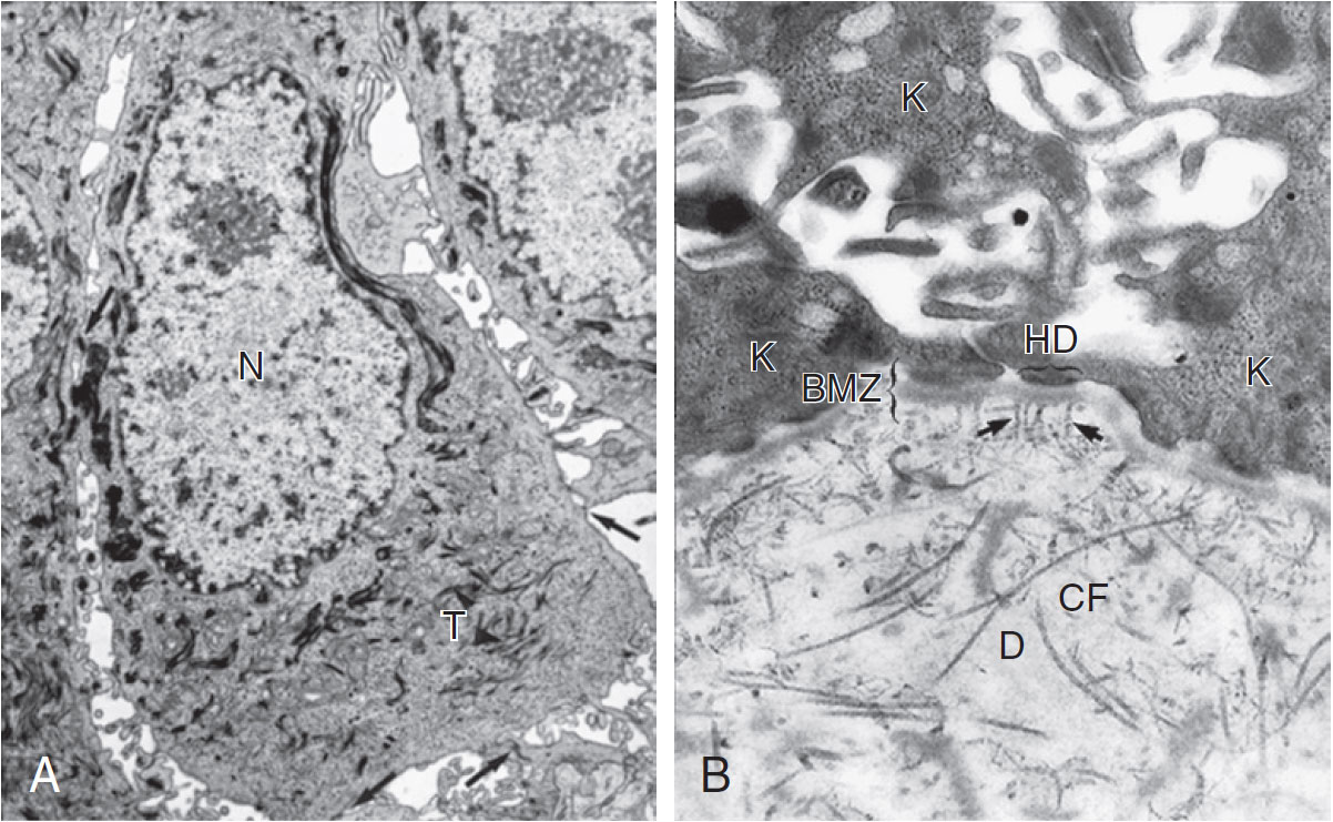
Describe the structure of the basement membrane zone (BMZ). | Fig. 1.3 A, Keratinocyte. An electron micrograph illustrates the ultrastructural components of a typical keratinocyte in the stratum spinosum, including the nucleus (N), tonofilaments (T), and desmosomal intercellular connections (arrow) that give this layer its 'spiny' appearance.
B, Basement membrane zone (BMZ). At the interface of the basal keratinocytes (K) of the epidermis and dermis (D) is the BMZ. The keratinocytes are attached to the BMZ by hemidesmosomes (HD). The BMZ is composed of the lamina lucida, which is the upper clear area, and the lamina densa, which is the dark area just below the lamina lucida. Anchoring fibrils (arrows) bind the BMZ to the dermis by intercalating among the collagen fibers (CF) of the dermis. | The BMZ is not normally visible by light microscopy in sections stained with hematoxylin-eosin but can be visualized as a homogeneous band measuring 0.5 to 1.0 mm thick on periodic acid–Schiff staining. Ultrastructural studies and immunologic mapping demonstrate that the BMZ is an extremely complex structure consisting of many components that function to attach the basal cell layer to the dermis (Fig. 1-3B). Uppermost in the BMZ are the cytoplasmic tonofilaments of the basal cells, which attach to the basal plasma membrane of the cells at the hemidesmosome. The hemidesmosome is attached to the lamina lucida and lamina densa of the BMZ via anchoring filaments. The BMZ, in turn, is anchored to the dermis by anchoring fibrils that intercalate among the collagen fibers of the dermis and secure the BMZ to the dermis. The importance of these structures in maintaining skin integrity is demonstrated by diseases such as epidermolysis bullosa, in which they are congenitally missing or damaged. |