|
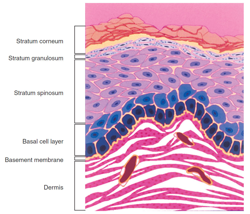
| Fig. 1.2 Epidermal layers and papillary dermis. |
The epidermis has four layers: the basal cell layer, spiny cell layer, granular cell layer, and cornified layer (Fig. 1-2). The basal cell layer (
stratum basalis) is composed of columnar or cuboidal cells that are in direct contact with the basement membrane, the structure that separates the dermis from the epidermis. The basal cell layer contains the germinative cells, and, for this reason, occasional mitoses may be present.
|
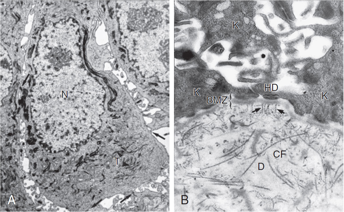
| Fig. 1.3 A, Keratinocyte. An electron micrograph illustrates the ultrastructural components of a typical keratinocyte in the stratum spinosum, including the nucleus (N), tonofilaments (T), and desmosomal intercellular connections (arrow) that give this layer its 'spiny' appearance. B, Basement membrane zone (BMZ). At the interface of the basal keratinocytes (K) of the epidermis and dermis (D) is the BMZ. The keratinocytes are attached to the BMZ by hemidesmosomes (HD). The BMZ is composed of the lamina lucida, which is the upper clear area, and the lamina densa, which is the dark area just below the lamina lucida. Anchoring fibrils (arrows) bind the BMZ to the dermis by intercalating among the collagen fibers (CF) of the dermis. |
The three layers above the basal cell layer are histologically distinct and demonstrate differentiation of the keratinocytes as they move toward the skin surface and become “cornified.” Just above the basal cell layer is the piny cell layer (
stratum spinosum), so called because of a high concentration of desmosomes and keratin filaments that give the cells a characteristic “spiny” appearance (Fig. 1-3A). Above the spiny layer is the granular cell layer (
stratum granulosum). In this layer, keratohyalin granules are formed and bind to the keratin filaments (tonofilaments) to form large electron-dense masses within the cytoplasm that give this layer its “granular” appearance.
The outermost layer is the cornified layer (
stratum corneum), where the keratinocytes abruptly lose all of their organelles and nuclei. The keratin filaments and keratohyalin granules form an amorphous mass within the keratinocytes, which become elongated and flattened, forming a lamellar array of “corneocytes.” The corneocytes are held together by the remnants of the desmosomes (dense bodies) and a “cementing substance” released into the intracellular space from organelles called Odland bodies.