How do atrophy and lichenification differ? |
|
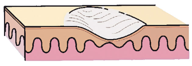
| Fig. 2.5 Atrophy. |
 |
| Fig. 2.7 Lichen simplex chronicus. Patient with atopic dermatitis and secondary lichenification manifesting as thickened skin with accentuation of skin markings. Secondary excoriations are also present. (Courtesy of the Fitzsimons Army Medical Center teaching files.) |
Atrophy (Fig. 2-5) is thinning of the epidermis, dermis, or subcutis (fat). Epidermal atrophy leads to a fine, cigarette-paper wrinkling of the skin surface, whereas dermal and fat atrophy cause a depression in the skin
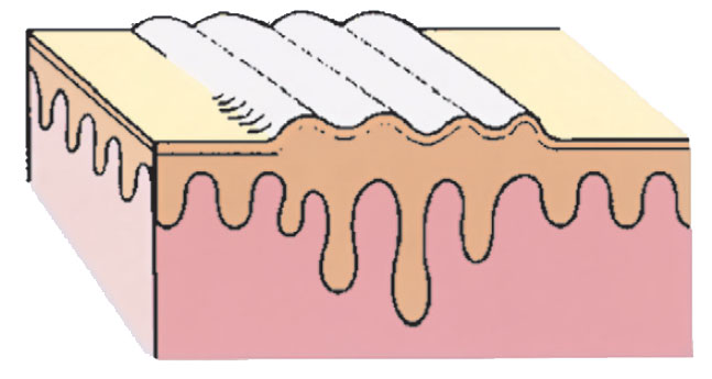 |
| Fig. 2.6 Lichenification. |
© 2025 Skin Disease & Care | All Rights Reserved.